Bladder cancer has one of the highest incidence rates in the world and ranks as the fourth most common tumour in men.

London: A team of Spanish researchers has successfully reduced the size of bladder tumours in mice by 90 per cent through a single dose of urea-powered nanorobots.
Bladder cancer has one of the highest incidence rates in the world and ranks as the fourth most common tumour in men.
Despite its relatively low mortality rate, nearly half of bladder tumours resurface within 5 years, requiring ongoing patient monitoring.
Frequent hospital visits and the need for repeat treatments contribute to making this type of cancer one of the most expensive to cure.
While current treatments involving direct drug administration into the bladder show good survival rates, their therapeutic efficacy remains low.
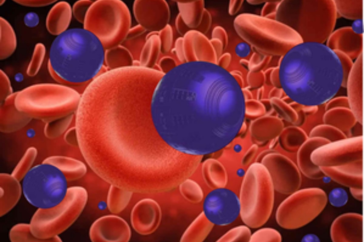
In the study, published in the journal Nature Nanotechnology researchers used nanorobots — tiny nanomachines consisting of a porous sphere made of silica. Their surfaces carry various components with specific functions.
Among them is the enzyme urease, a protein that reacts with urea found in urine, enabling the nanoparticle to propel itself. Another crucial component is radioactive iodine, a radioisotope commonly used for the localised treatment of tumours.
These advancements aim to reduce the length of hospitalisation, thereby implying lower costs and enhanced comfort for patients, said the team led by those from the Institute for Bioengineering of Catalonia (IBEC) and CIC biomaGUNE in collaboration with the Institute for Research in Biomedicine (IRB Barcelona) and the Autonomous University of Barcelona (UAB) in Spain.
“With a single dose, we observed a 90 per cent decrease in tumour volume. This is significantly more efficient given that patients with this type of tumour typically have 6 to 14 hospital appointments with current treatments. Such a treatment approach would enhance efficiency, reducing the length of hospitalisation and treatment costs,” said Samuel Sanchez, ICREA research professor at IBEC and leader of the study.
The next step, which is already underway, is to determine whether these tumours recur after treatment.
In previous research, the scientists confirmed that the self-propulsion capacity of nanorobots allowed them to reach all bladder walls.
This new study goes further by demonstrating not only the mobility of nanoparticles in the bladder but also their specific accumulation in the tumour. This achievement was made possible by various techniques, including medical positron emission tomography (PET) imaging of the mice, as well as microscopy images of the tissues removed after completion of the study.
The latter were captured using a fluorescence microscopy system developed specifically for this project at IRB Barcelona. The system scans the different layers of the bladder and provides a 3D reconstruction, thereby enabling observation of the entire organ.

