by Light Publishing Center, Changchun Institute of Optics, Fine Mechanics And Physics, CAS

The detection of individual particles and molecules has opened new horizons in analytical chemistry, cellular imaging, nanomaterials, and biomedical diagnostics. Traditional single-molecule detection methods rely heavily on fluorescence techniques, which require labeling of the target molecules.
In contrast, photothermal microscopy has emerged as a promising label-free, non-invasive imaging technique. This method measures localized variations in the refractive index of a sample’s surroundings, resulting from light absorption by sample components, which induces temperature changes in the surrounding region. Whispering gallery mode (WGM) resonators are ultra-sensitive temperature sensors due to their ultra-high quality (Q) factors.
In a new paper published in Light: Science & Applications, a research group led by Prof. Judith Su from the Wyant College of Optical Sciences and the Department of Biomedical Engineering at the University of Arizona demonstrated label-free ultra-sensitive photothermal microscopy using microtoroid WGM optical resonators.
This technique enables the detection of single nanoparticles as small as 5 nm quantum dots with a signal-to-noise ratio (SNR) exceeding 10,000. This method significantly enhances photothermal sensitivity, achieving a detection limit of 0.75 pW in heat dissipation. This advancement allows for the accurate detection of single molecules, showcasing the capabilities of photothermal microscopy.
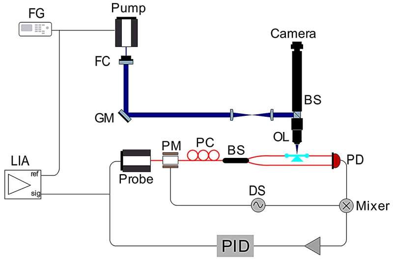
Su’s group previously developed a system called Frequency Locked Optical Whispering Evanescent Resonator (FLOWER), which uses frequency locking to track the resonance shift of optical microcavities. This photothermal microscopy system employs FLOWER to measure the photothermal signal. The resonance shift, excited by a free-space pump laser, is measured by the system.
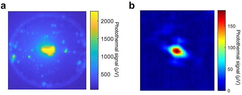
The pump laser, with a 203.7 Hz amplitude modulation (AM), illuminates the microtoroid through a galvo mirror scan system. The resulting 203.7 Hz oscillation of the resonance shift is detected by FLOWER. A lock-in amplifier is used to measure the amplitude of the resonance shift oscillation. A 2D spatial scan of the pump laser generates a photothermal image of the microtoroid, with high photothermal spots used to detect single nanoparticles.

The researchers envision the potential of photothermal microscopy and say, “Moreover, future advancements may involve spectroscopy measurements by varying the pump laser’s wavelength or exciting with different wavelengths to enable multicolor imaging. In future work, although not necessary for many applications, we can also combine this system with specific capture agents such as aptamers or sorbent polymers to provide enhanced specificity.
“The integration of photothermal microscopy with FLOWER opens possibilities for real-time observation of dynamic changes and interactions of target molecules. We believe that overall, FLOWER based photothermal microscopy represents a versatile platform for label-free imaging and single-molecule detection. The demonstrated high sensitivity and discrimination capabilities pave the way for advancements in nanoscale imaging and characterization techniques.”
More information:
Shuang Hao et al, Single 5-nm quantum dot detection via microtoroid optical resonator photothermal microscopy, Light: Science & Applications (2024). DOI: 10.1038/s41377-024-01536-9
Provided by
Light Publishing Center, Changchun Institute of Optics, Fine Mechanics And Physics, CAS
Citation:
Ultra-sensitive photothermal microscopy technique detects single nanoparticles as small as 5 nm (2024, August 23)
retrieved 23 August 2024
from https://phys.org/news/2024-08-ultra-sensitive-photothermal-microscopy-technique.html
This document is subject to copyright. Apart from any fair dealing for the purpose of private study or research, no
part may be reproduced without the written permission. The content is provided for information purposes only.

